
Autism – the Th17–IL-17–IL-17R System
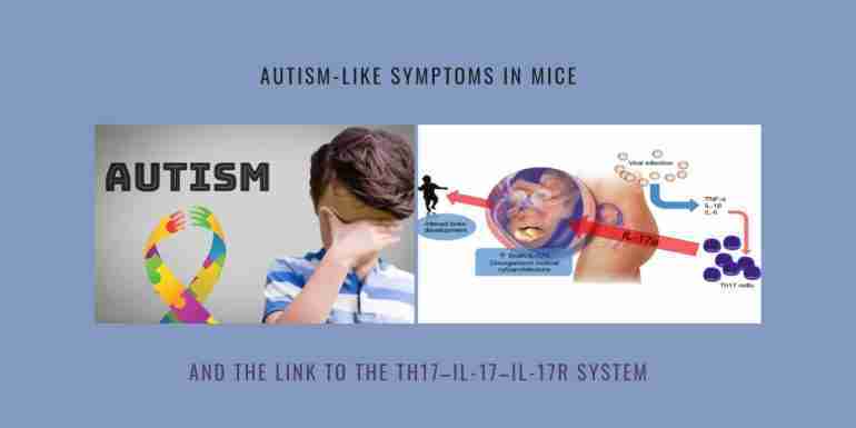
 A new report published in Science indicates that the key pro-inflammatory cytokine interleukin (IL)-17a may interfere with the normal brain development in a mouse model of autism, causing disorganized cortical cytoarchitecture and autism-like symptoms.
Previous research also suggests that viral infection of mothers during pregnancy may contribute to a higher incidence of ASD in the offspring (Atladóttir et al., 2010; Lee et al., 2015). Several studies have suggested this link.
Thus, in the rodent maternal immune activation (MIA) model of this phenomenon, offspring from pregnant mice infected with virus or injected intraperitoneally with synthetic double-stranded RNA (dsRNA) [poly(I:C)], a mimic of viral infection, exhibit behavioral symptoms reminiscent of ASD: social deficits, abnormal communication, and repetitive behaviors – and autism-like symptoms.
IL-17 is a highly pathogenic pro-inflammatory cytokine, considered a major player in several autoimmune/inflammatory diseases, whereas altered cytokine profiles and increased serum IL-17 levels.
TH17 cells and their cytokine mediators have been suggested to have a role in ASD. For example, elevated levels of interleukin-17a (IL-17a), the predominant TH17 cytokine, have been detected in the serum of a subset of autistic children. A genome-wide copy number variant (CNV) analysis identified IL17A as one of many genes enriched in autistic patients. Similarly, in the MIA mouse model, CD4+ T lymphocytes from affected offspring produced higher levels of IL-17a upon in vitro activation.
A 2012 study indicates that children with autism have significantly higher serum IL-17A levels than healthy controls. Of note, the levels correlated significantly with the severity of autism: Patients with severe autism had significantly higher serum IL-17A levels than those with mild to moderate autism and raised serum IL-17A levels were significantly more common in children with severe autism. This is perhaps the first study to measure levels of IL-17A in relation to the severity of autism.
However, no study has yet correlated the Th17 cell responses in pregnant women with the risk of ASD development in the offspring.
A disease model in rodents, referred to as ‘maternal immune activation’ (MIA) has been often used to study viral and immune activation in the mother during pregnancy, and the link to autism-like behaviors in the offspring. In this model pregnant mice are injected with the synthetic double-stranded RNA, poly(I:C), to mimic a viral infection.
Previous data indicate that in the MIA model, with autism-like symptoms, the immune cells, and particularly the CD4+ T lymphocytes from the affected offspring, produce high amounts of IL-6 and IL-17 upon in vitro stimulation. In addition, earlier studies showed that MIA induces in the offspring behavioral impairments similar to those that are observed in autistic children. This includes reduced social interaction, abnormal communication and stereotyped/repetitive behaviour.
In the Science report, Gloria Choi and colleagues from the Massachusetts Institute of Technology, Cambridge, MA, and the New York University School of Medicine, NY used the MIA model to study if the highly pathogenic IL-17-IL-17R pathway may contribute to abnormal fetal brain development and autism-like behavioral phenotypes.
The authors found that in mothers, T helper (Th)17 cells and their effector cytokine IL-17 are required before the MIA-induced behavioral abnormalities (and autism-like symptoms) are observed in the offspring.
They report that pregnant mothers, injected with poly(I:C), had a strong induction of serum IL-6 and IL-17 cytokine levels (of note, IL-6 is a key factor for Th17 cell differentiation). In addition, they found that the expression of the IL-17 receptor subunit A (IL-17Ra) was strongly augmented in the fetal brain upon induction of MIA.
Importantly, the maternal IL-17a was able to induce abnormal cortical development in offspring, related to patches of disorganized cortical cytoarchitecture. Moreover, the maternal IL-17a promoted ASD-like behavioral abnormalities in the offspring.
Corroborating this data, in the study of Choi et al., the injection of pregnant mothers with antibodies against IL-17 supressed the increased IL-17Ra expression in fetus brain and prevented the development of ASD-like behavioral abnormalities.
Thus, these results suggest that an immunological activation during pregnancy may have important long-term outcomes in the offspring. This study finds that the Th17-IL-17-IL-17R pathway is able to affect neuronal development, and, identifies a specific maternal immune cell population that exerts a direct role in inducing ASD-like phenotypes by acting on the developing fetal brain.
Of note, the authors discuss that some of the IL-17 effects might be related to the structural similarities detected between the IL-17 family cytokines and neurotrophins such as the nerve growth factor.
Also of interest is a recent study, published in Cell, indicating that Th17 cells are able to induce tertiary lymphoid tissues (TLTs) within the brain meninges, which are linked to the formation of an immune-competent stromal cell niche in the meninges and local demyelination.
Source: Science, 2016, 351:933-9. doi: 10.1126/science.aad0314. Epub 2016 Jan 28.
Read More: science.sciencemag.org
Updates
2020
A 2020 article entitled: ‘Aberrant IL-17 Levels in Rodent Models of Autism Spectrum Disorder: A Systematic Review’ is perhaps the first systematic review assessing IL-17 levels in rodent models of Autism spectrum disorder (ASD). The review shows that IL-17 levels are increased in different tissues and in the serum of ASD rodent models, as reported by 23 out of 28 studies included in this review. Also, increased IL-17 levels could be observed in pregnant mice, embryos, and offspring, highlighting the possible role of this cytokine during neurodevelopment, pathogenesis and pathophysiology of ASD, as well as in behavioural alterations.
2022
Another 2022 systematic review reports increased IL-17 levels in the serum of subjects with autism. Of note, this review concludes that neutrophils and CD4+ T cells are perhaps the potential sources of IL-17 in the steady state of autism, in addition to IL-17 production by stimulated CD4+ T cells.
As per this review, neutrophils of individuals with autism express the IL-17RC subunit, which is absent in the neutrophils of control subjects. This enables neutrophil to potently respond to IL-17A/IL-17R signaling and release IL-6, leading to increased IL-17 production in autism. Thus, taken together, these results suggest that Th17 cells and neutrophils are potential sources of upregulated IL-17 levels in autism.
As per another 2022 review, during pregnancy, the placenta’s regulatory and pro-inflammatory T-cell activity must be carefully coordinated. Uterine immune activation (UIA) alters the cytokine milieu, increasing IL-6 levels, to favor the activation of maternal Th17 cells. Furthermore, UIA at E12.5 enhanced the abundance of the uterine hybrid CD4+CD25+ Foxp3+RORγT+Tregs (Tregs-Th17), indicating a pathological transition of Tregs toward the Tregs–Th17 phenotype and the generation of hybrid Tregs capable of secreting IL17a.
2024
A 2024 study by Dina E. Sallam et al. published in BMC Pediatrics found that the serum levels of IL-17 A and IL-22 were significantly higher in autism spectrum disorder (ASD) patients than in controls. Of note, however, only IL-22 had a significant positive correlation with ASD severity by Childhood Autism Rating Scale (CARS) scores.
Thus, according this study raised serum levels of IL-17 A and IL-22 are associated with ASD; but only IL-22, not IL-17 A, correlated with ASD severity.
Another 2024 study by Yehoshua Willinger et al. published in International Journal of Molecular Sciences reported an improved social behavior and reduced overall ASD-like behavior following by two injections of the cytokine IL-17A in valproic acid (VPA)-exposed mice.
The authors found that serum levels of Th17-related cytokines, i.e., IL-17A in males and IL-17F and IL-21 in females, were positively correlated with social behavior and negatively correlated with the total ASD score.
According to the authors their study suggested a complex role for IL-17A in ASD. Thus, while contributing to its pathology at the developmental stage, IL-17 may also promote the alleviation of behavioral deficits post-developmentally by promoting neuritogenesis and synaptogenesis in the dentate gyrus.
2025
A brand new 2025 study by Pierre Ellul et al. from the Sorbonne Université, INSERM, Paris, France claims that the maternal immune activation (MIA) during pregnancy imprints a Treg deficiency in offspring that perpetuates brain inflammation and an autism-like phenotype, which can be reversed by Treg stimulation.
Background: The causal relationship between MIA and autism spectrum disorder (ASD) is supported by experimental models. Poly(I:C) injection into pregnant mice, to induce MIA, triggers symptoms in the offspring that resemble core ASD symptoms in humans, including communication deficits, repetitive behaviors, and impaired social interactions. Mechanistically, the current view is that MIA disrupts neurodevelopment by activating maternal effector T helper (Th17) cells, which in turn produce interleukin-17a (IL-17A). This pro-inflammatory cytokine crosses the placenta and binds to IL-17A receptors on fetal neurons, thereby affecting neurodevelopment. The ASD phenotype is driven by long-term activation of the IL-17A pathway in the brain, which induces disruption of synaptic connectivity (for more information and references of these studies, see the introduction of this article).
Importantly, the authors also discuss that they established a causal link between Treg deficiency and ASD, by showing that Treg stimulation can normalize both the biological and the clinical symptoms in ASD-like young adult mice. According to these authors, this is significant because it shows that a Treg defect is not only important for initiating the autistic core symptoms during prenatal growth but is also involved in the maintenance of these symptoms in the adult offspring.
Related stories you may like:
Monocyte Chemotactic Protein-1 (MCP-1): High Neonatal Levels May Represent an Early Marker of Autism
Cortisol Diurnal Variation, IL-6 and TNF-alpha as Potential Diagnostic Markers in Autism
Cytokine Dysregulation Associated with Behavioral Deficits in a Mouse Model of Autism
Cytokine Profiles in Subjects with Autism Spectrum Disorders
Cytokine Profiles in Mothers Bearing Autistic Children
Prenatal Immune Challenge – ‘Disease Primer’ Increasing the Offspring’s Vulnerability to Stress